Research
Methods & Equipment
Methods
Experimental
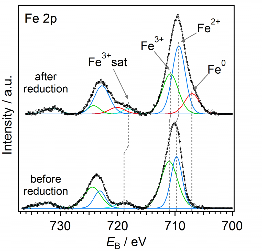
- in-situ Near Ambient Pressure X-Ray Photoemission Spectroscopy (in-situ NAP-XPS)
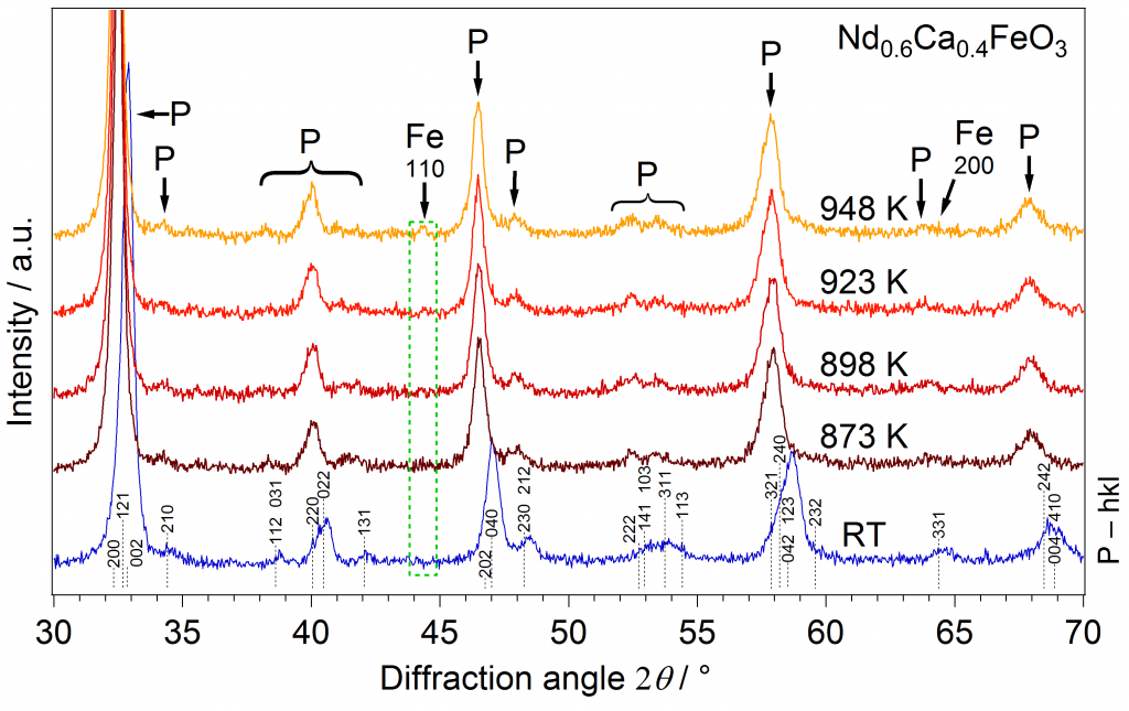
- (in-situ) X–Ray Diffraction (in-situ XRD) and X-Ray Absorption Spectroscopy (XAS)
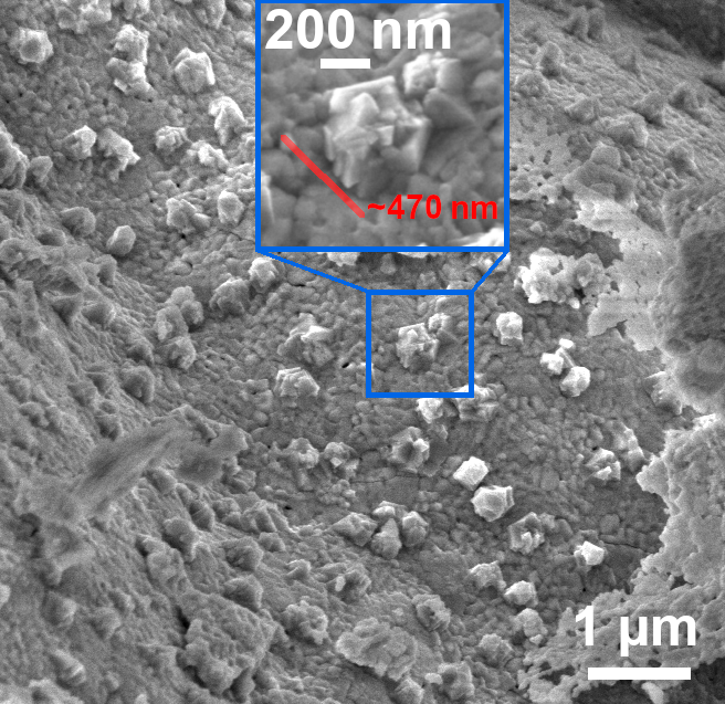
- Scanning Electron Microscopy (SEM) and Scanning Tunnelling Microscopy (STM)
- Gas Chromatography (GC) and Mass Spectrometry (MS)
- Low Energy Ion Scattering (LEIS) and Low Energy Electron Diffraction (LEED)
- Temperature Programmed Reduction (TPR) and Oxidation (TPO)
- Electrochemical Impedance Spectroscopy (EIS)
- Pulsed Laser Deposition (PLD), a version of Physical Vapour Deposition (PVD)
Theoretical
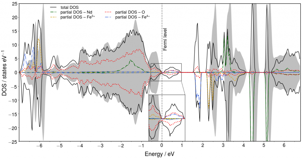
- Density Functional Theory (DFT), using the software package WIEN2k
Equipment...
... of the Group
Reactor for Catalytic Testing – Co-owned with ClusCAT Lab
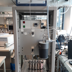
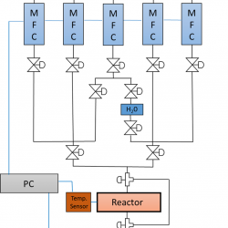
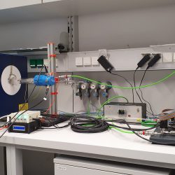
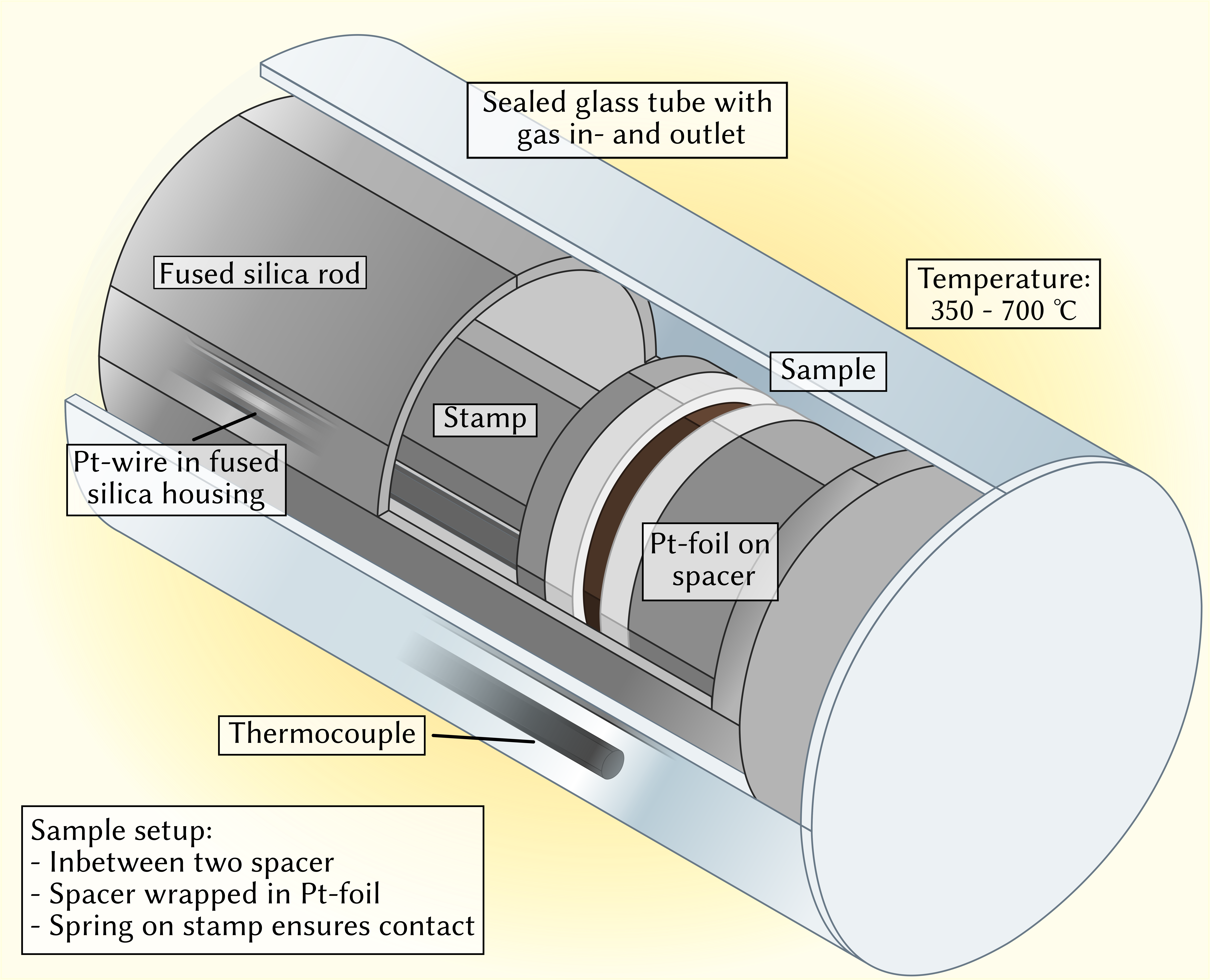
For the basic catalytic characterization of our materials, we use a custom-built fixed bed reactor coupled with a micro-GC system from Inficon. Normally, H2, CO, CO2, O2, CH4, and Ar are mounted onto the system, but the easily accessible Swagelok connections allow a simple and quick change to virtually any gas. The gases are mixed in a mass flow controller setup and can the be led through a saturator, allowing us to introduce water into the system.
After the oven, the micro-GC system is the heart of the setup. It analyses the gas on three different columns simultaneously. This gives us great versatility with respect to the compounds we want to analyse. From small molecules such as CO or H2 to bigger alkanes up to C4 or C5 nearly everthing is possible. And all that with a sensitivity down to the ‰ range.
Near Ambient Pressure X-Ray Photelectron Spectroscopy (NAP-XPS)
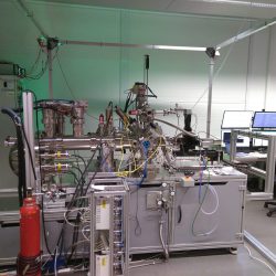
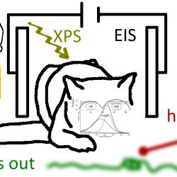
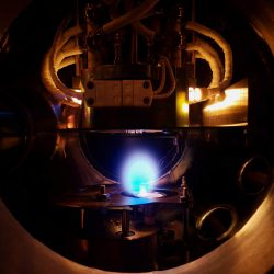
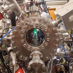
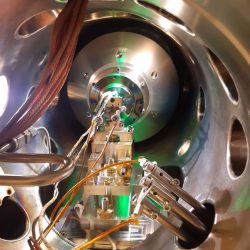
The flagship of the TUCAS project is the in-situ Near Ambient Pressure X-Ray Photoelectron Spectroscopy setup — we invested about 1 Mio. € to build it. A commercial XPS setup (by SPECS) was used as basis and modified and adapted to our needs: our setup combines XPS for surface investigation (oxidation state, elemental composition) with electrochemical impediance spectroscopy for bulk characterisation (ion-conductivity or ion-mobility) and mass spectrometry for gas phase analysis.
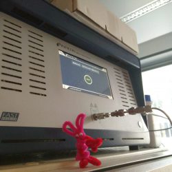
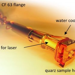
A specially designed custom-made sample stage allows both measurements to be conducted in-situ, i.e. under reaction conditions. We can perform reactions up to 1000 °C and pressures of several mbar. Reaction temperatures are reached with direct laser heating, and the temperature control is handled in different ways: directly via installed thermocouple or pyrometer, or indirectly via resitivity measurements during impedance spectroscopy. We can realize different gas atmospheres with a gas mixing setup consisting of multiple massflow controllers — gas phase analysis is done with a Prism Pro Mass Spectrometer from Pfeifer.
... available at TU Wien
Electrochemical Impedance Spectroscopy (EIS) Setup – Designed by Huber Scientific
Pulsed Laser Deposition (PLD) Chamber
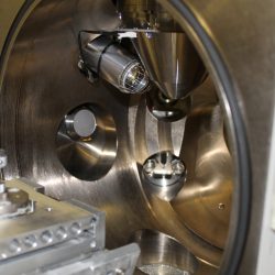
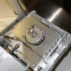
We employ Pulsed Laser Deposition (PLD) as conventional PVD technique to obtain geometrically well-defined thin films for EIS measurements. Most of our devices are custom-built and this high vacuum PLD chamber is no exception.
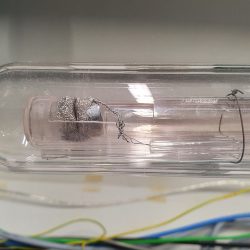
The necessary high vacuum is achieved via a turbo pump from Leybold. The deposition is accomplished via a Kr/F excimer laser (COMPex Pro 201F) by ablating a self-rotating high purity target placed in a target holder mounted on a target carousel. A fully motorised substrate holder allows easy sample switching as well as accurate tuning of the target-substrate distance. An also motorised IR-heater is used for mostly uniform heating of the substrate surface. The desired background gas atmosphere can be achieved via a simple mass flow controller setup.
SEM/TEM: Electron Microscopy at USTEM
For microscopy, we profit from the wide variety of instruments and expertise available at the microscopy service center of TU Wien (USTEM — Universitäre Service-Einrichtung für Transmissions-Elektronenmikroskopie).
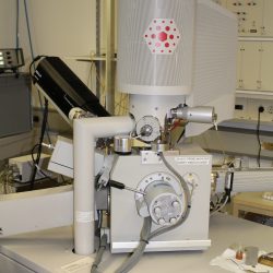
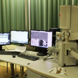
There, we can perform scanning electron microscopy (SEM, FEI Quanta 250 FEGSEM microscope) or transmission electron microscopy (TEM, FEI Tecnai F20 microscope), possibly combined with spatially resolved elemental analysis by Energy Dispersive X-Ray spectroscopy (EDX). Furthermore, advanced options for sample preparation are possible, such as cutting out lamellas with a focused Ga ion beam (FIB, executed in a FEI Quanta 200 3D DBFIB).
These techniques open up the nanoscale regime, offering a direct view of our samples with down to atomic resolution. By revealing their detailed surface morphology and composition, this complements our other methods and helps us to understand what is going on. Thus, microscopy is an integral part of our materials characterization – and, what’s more, yields beautiful images.
X-Ray Diffraction (XRD) at the X-Ray Center
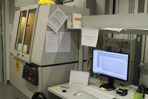
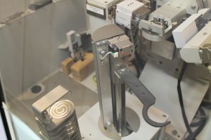
Our X–Ray Diffraction (XRD) measurements for structural characterization are carried out at the TU Wien X-Ray Center. They provide PANalytical X’Pert Pro diffractometers with Cuα1,2 radiation and an X’Celerator linear detector, suitable for powder diffraction experiments in Bragg-Brentano geometry. These instruments enable quick analysis with minimal preparation and high flexibility with respect to the nature of samples.
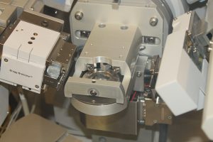
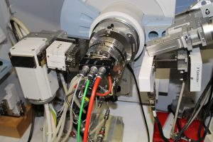
Furthermore, we can perform in-situ or operando XRD experiments at the X-Ray Center, using an Anton Paar XRK 900 chamber, where the measurement takes place while the sample is in a gas flow atmosphere at elevated temperatures. In combination with a custom-built mass flow controller setup to adjust the atmospheric composition and an optional post-chamber gas phase analysis with a mass spectrometer, we can get deep insights into the behavior of our materials during catalytic reaction, correlating structure and catalytic performance.
... at (inter)national Research Centers
Synchrotron Facilities
The most promising of our samples — which we identify via lab measurements — are studied in even more detail at snychrotron facilities.
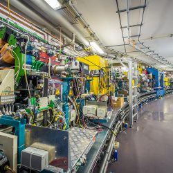
Synchrotrons are powerful analysis tools as they provide not only the possibility of using radiation of different wave lenghts, but also make high-intensity radiation available. Thus, high resolution data can be obtained (due to better signal-to-noise ration and narrower line widths). Moreover, we can apply larger pressures during experiments (due to higher intensities) and do depth profiles (with different wave lenghts).
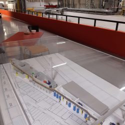
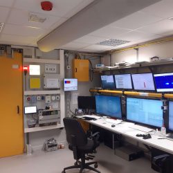
In the past, we have used the ISISS Station of BESSY II at the Helmholtz Zentrum Berlin, Germany; the HAXPES Spectrometer (more specificially the Polaris instrument) and the HEMS beamline (HR-SXRD) of the PETRA III radiation source at the DESY in Hamburg, Germany; and the HIPPIE beamline of MAX IV Laboratory in Lund, Sweden.
Vienna Scientific Cluster
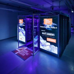
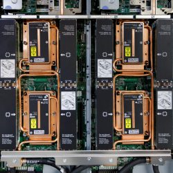
We use the 4th generation of the Vienna Scientific Cluster (VSC4) for our theoretical calculations.
VSC4 is the most powerful supercomputer ever built in Austria – at its launch in June 2019, it was ranked 82nd in the global Top500 list of supercomputers. It boasts a peak performance of 2.7 petaflops (one petaflop is a quadrillion – a million billion – operations per second).
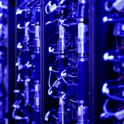
790 watercooled nodes with 48 cores each (at 3.1 GHz per core) result in a total number of 37,920 cores and each node provides at least 96 GB RAM (there are nodes with 388 and 768 GB available as well). The total storage capacity of is a whopping 7 PB (i. e. 7 million GB).
Its predecessor VSC3 is also still available and expected to be in operation at least until the end of 2021.
